Patient assessment
1. Clinical examination
Hand surgery involves a multi-specialty approach to the assessment, treatment, and aftercare of hand trauma. Therefore, the specialized teams should be involved early on according to the specific injury requirements.
The clinical signs of wrist injuries include swelling, pain, and tenderness over the injured bone.
Swelling and hematoma are more typical for complex injuries with fracture-dislocation.
In simple fractures, eg, of the scaphoid waist, swelling and hematoma are not typical. A problem with simple fractures is that many patients seek medical care late because they have only minimal symptoms.
Assess the range of motion of the wrist in all directions.
Note any painful range of motion restrictions.
For a suspected scaphoid fracture, check the anatomic snuff box for tenderness.
For a suspected established scapholunate injury, perform the Kirk-Watson test.
- Watson HK, Ashmead D, Makhlouf MV. Examination of the scaphoid. J Hand Surg. 1988;13A:657–660.
Ask the patient for numbness of the thumb to the ring finger to assess the possibility of carpal tunnel syndrome. This frequently occurs with complex injuries, eg, greater and lesser arc injuries, and needs acute surgery or preliminary closed reduction and splinting for delayed (but early) surgical management.
2. Radiological assessment
Introduction
X-rays form the basis of assessment of carpal fractures and dislocations.
Further definition of complex fractures and dislocations is facilitated by CT scanning, eg, if a fracture of the scaphoid waist is suspected but not clearly visible on x-ray. The role of MRI in carpal injuries is less well defined.
Radiological signs in the carpal bones
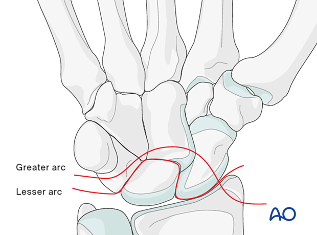
‘Arcs’ are lines that can be drawn or imagined on x-ray/CT images of the hand and wrist to help assess the alignment of the carpal bones. A discontinuity in an arc indicates a malalignment of the carpal bones either by the fracture or dislocation and should lead to further investigation, eg, CT scan.
Variations of injury patterns can be identified depending on which carpal bones and ligaments are affected and the direction of any dislocation or fracture displacement.
Greater arc injuries comprise fracture-dislocations of the scaphoid, capitate, hamate, and/or triquetrum.
Lesser arc injuries are pure ligamentous injuries around the lunate.
The concept of ‘arcs’ helps to identify the location and extent of a complex carpal injury.
X-ray imaging
Standard wrist x-rays include PA, lateral, and oblique views.
In this case, the AP and lateral x-rays show a fracture line across the waist of the scaphoid with normal carpal alignment.
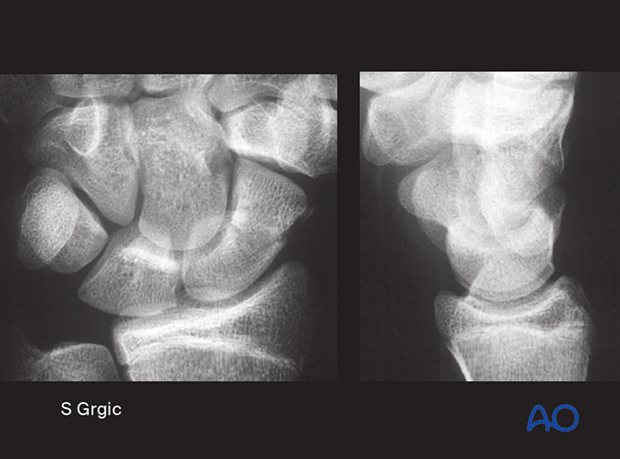
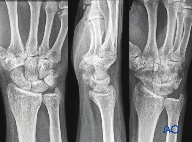
This case shows x-rays of a lunate dislocation. This injury is difficult to identify on an x-ray AP view. In the lateral view, the so-called spilled-teacup sign is clearly visible.
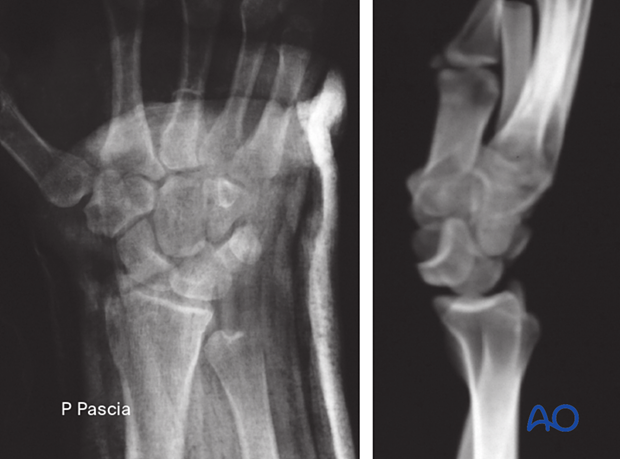
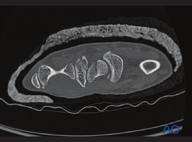
In cases of so-called “occult” fractures, the fracture is suspected clinically but not visible on the x-rays.
A CT scan may then be helpful.
This case shows a scaphoid waist fracture.
Stress x-ray imaging
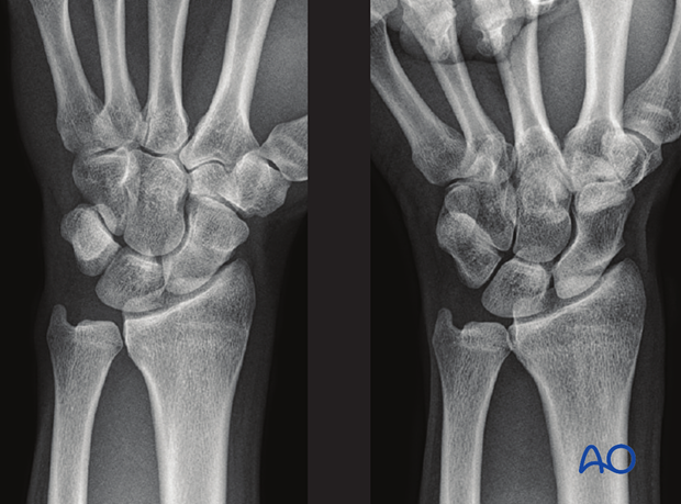
In suspected scapholunate injuries, standard x-rays are usually insufficient to diagnose the condition correctly. A stress x-ray can help to demonstrate dynamic wrist instability.
In this case, the x-ray with a clenched fist (right image) shows a gap at the scapholunate junction indicating a scapholunate ligament injury with dissociation.
CT and MRI
In an apparently isolated fracture, associated carpal instability can be difficult to identify but should be suspected. If available, CT imaging may be helpful.
MRI is preferred to define the presence of ischemia as a consequence of a fracture.
In this case, the 2-D CT scan shows a fracture with minimal displacement through both cortices of the scaphoid in the sagittal plane.
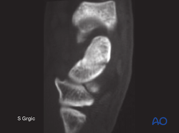
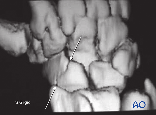
The 3-D CT renderings show the fracture and its relation to the lunate and capitate in a case of suspected scapholunate fracture-dislocation. The arrows indicate the scaphoid fracture from the ventral …
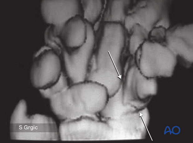
… and dorsal aspects.
