Neurological anatomy, protection, and handling
1. Introduction
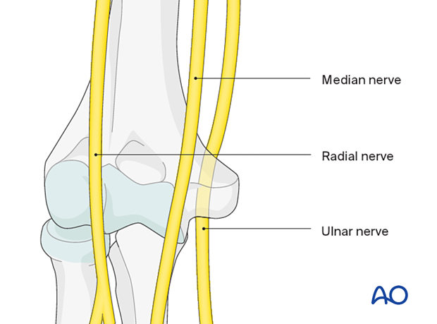
All nerves crossing the elbow are at risk in elbow fractures and surgical approaches to fix them. The most common nerve injuries are related to the radial and ulnar nerve.
2. Radial nerve
The radial nerve is at risk:
- In extraarticular fractures
- During retraction to expose the lateral column of the humerus
Relevant applied anatomy
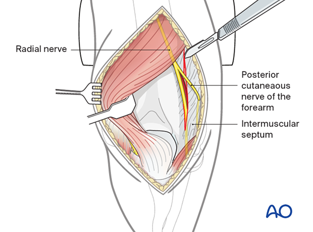
The radial nerve passes from the posterior aspect of the humerus into the anterior compartment of the arm through an opening of the lateral intermuscular septum at the junction of the upper 3/5 and lower 2/5 of the lateral side of the humerus.
The radial nerve is relatively fixed at this transition point.
It is also relatively fixed where it bifurcates immediately distal to the elbow in the forearm.
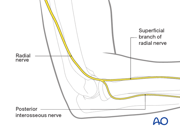
The working length of the radial nerve is the distance between these two points. Take care when applying retractors in this area as the nerve may be stretched.
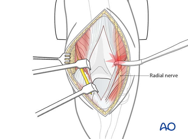
Risk reduction
To reduce the risk of nerve stretching during fracture exposure, the lateral intermuscular septum may need to be incised.
3. Ulnar nerve
The ulnar nerve is at risk:
- In distal metaphyseal and intraarticular fractures of the distal humerus
- During exposure and retraction of soft tissues around the medial column
- During insertion of K-wires into the articular block from the medial side
Relevant applied anatomy
The ulnar nerve passes from the anterior compartment of the arm into the posterior compartment through an opening of the medial intermuscular septum at the junction of the upper 3/5 and lower 2/5 of the medial side of the humerus.
The ulnar nerve is relatively fixed at this transition point.
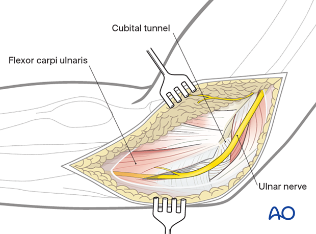
The ulnar nerve is also relatively fixed where it enters the flexor carpi ulnaris muscle immediately distal to the elbow in the forearm.
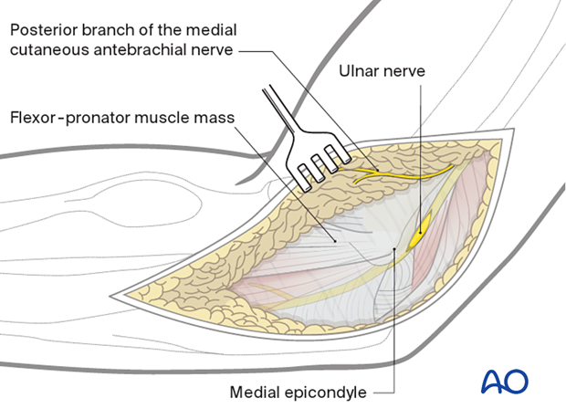
The working length of the ulnar nerve is the distance between these two points. Take care when manipulating bone fragments or applying retractors in this area as the nerve may be stretched.
Since the nerve is relatively fixed close to the elbow joint, it is at much greater risk of injury by both the fracture and surgical approach than the radial nerve.
Risk reduction
To reduce the risk of injury to the ulnar nerve:
- The position of the nerve should be monitored throughout the entire procedure
- The medial intermuscular septum may be incised
- The ulnar nerve may be mobilized from the cubital tunnel and released by partial incision of the flexor carpi ulnaris
4. Median nerve
The median nerve is at risk:
- In anteriorly displaced fractures of the distal humerus
- Fractures with associated vascular injury
- Insertion of K-wires into the articular block
Relevant applied anatomy
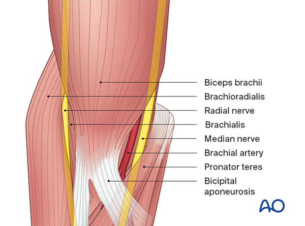
The median nerve crosses the anterior capsule of the elbow joint, running into the forearm between the two heads of the pronator teres.
