Approaches to the radius for intramedullary nailing
1. Introduction
The correct nail entry point and so the chosen approach depends on the nail type used. The following entry points are used:
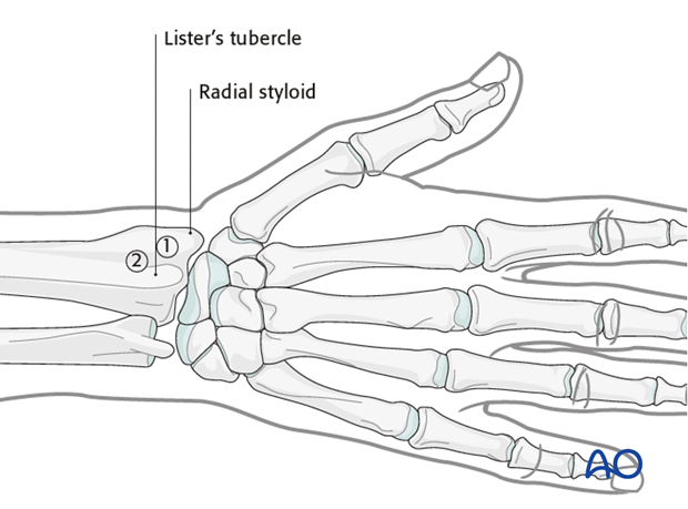
- On the ulnar side of the radial styloid, between the first and second extensor tendon compartments
- On the radial side of the Lister’s tubercle, between the second and third extensor tendon compartments
In the following, we shall describe the approach to the radial side of the Lister's tubercle (2). For other approaches, please refer to the manual of your chosen nail system.

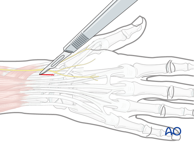
2. Skin incision
Make a 2.5 to 3 cm longitudinal skin incision over the distal radius on the radial side of Lister’s tubercle.

3. Deep dissection
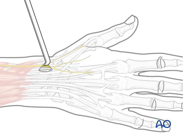
Bluntly dissect subcutaneous tissues to avoid injury to the dorsal branches of the superficial radial nerve, which cross the tendons closely.
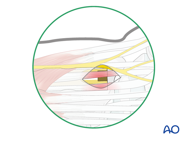
A skin incision is made and then progressively deepened by spreading a forceps and advancing the retractors layer by layer until the bone is reached. It may be necessary to incise a portion of the extensor retinaculum.

Pitfalls
Any entry point near the Lister’s tubercle can damage the extensor tendons of the second and third compartments, especially when the nail is prominent.
Complete division of the extensor retinaculum can result in tendon subluxation (“bowstringing”).


