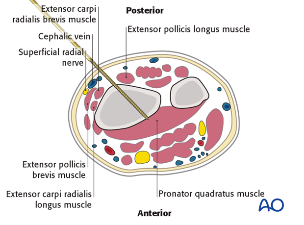
Safe zonesin the forearm for pin placement
1. Anatomical considerations
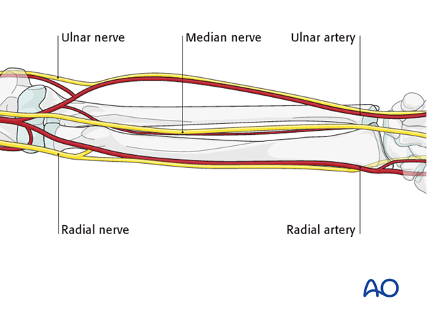
The forearm anatomy is complex due to the presence of three major neurovascular bundles.
In particular:
- Radial nerve with its two branches (i.e., posterior interosseous nerve and superficial radial nerve)
- Radial artery
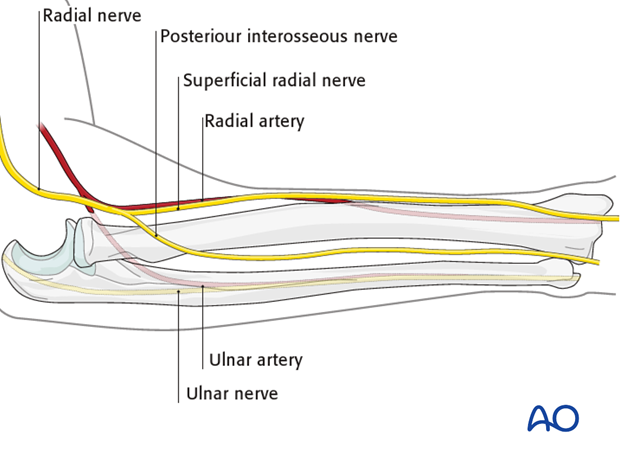
- Median nerve
- Ulnar nerve
- Ulnar artery
Except for the radial nerve, all previously mentioned structures run in the flexor compartment anteriorly.
Note: the cephalic vein runs parallel to the superficial radial nerve in the distal part of the forearm.
Pitfall: The more proximal the surgical field, the greater the risk of damage to important structures!
Usually pin insertion is via the posterolateral aspect of the radius. Avoid anterior pin insertion.
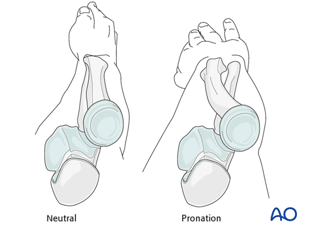
Forearm position
The anatomical relationships are complex because they depend on the rotational orientation. For pin insertion, two forearm positions are most often used:
- Pronation – for the ulna
- Neutral rotation (between pronation and supination) – for the radius
Skin tension
Pins should be inserted at sites where the soft-tissue envelope is as thin as possible. Moreover, the pins should be inserted between muscle compartments, not through them.
2. Safe zones in the ulna (pronation)
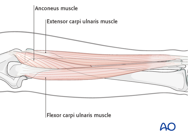
Safe zones for ulnar pin insertion are along the whole shaft between the extensor carpi ulnaris and the flexor carpi ulnaris.
3. Safe zones in the ulna
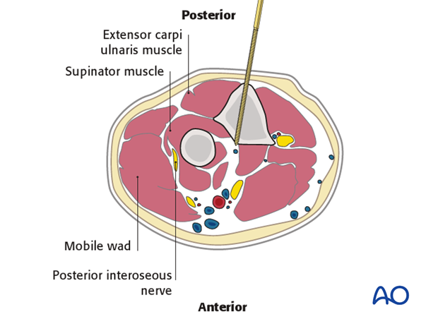
This cross section illustrates a pin inserted into the proximal ulna.
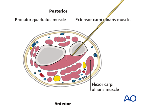
This illustration shows a pin inserted into the distal ulna.
4. Safe zones in the proximal radius
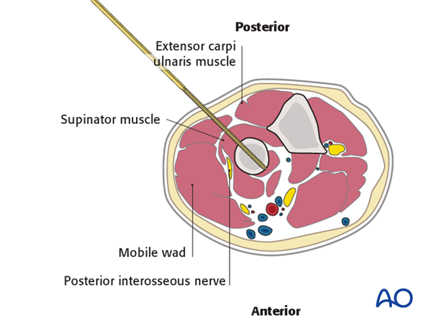
The radial nerve is vulnerable in this zone. The supinator muscle cloaks the dorsal aspect of the upper 3rd of the radius, with the posterior interosseous nerve running through its substance.
A skin incision is made and then progressively deepened by spreading a forceps and advancing the retractors layer by layer until the bone is reached.
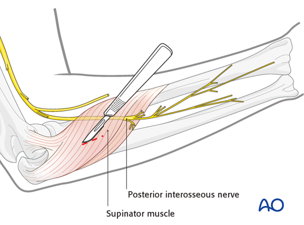
This cross section illustrates a pin inserted into the proximal third of the radius.
Beware of the posterior interosseous nerve!
5. Safe zones in the distal radius
Beware of the superficial radial nerve and outcropping muscles or extensor tendons.
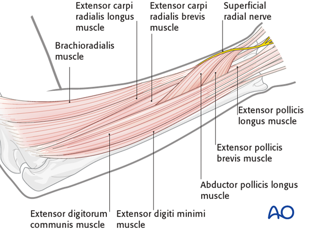
This illustration shows the distal third of the forearm. Any pins need to be inserted under direct vision using retractors down to the bone to avoid the superficial branch of the radial nerve.
A skin incision is made and then progressively deepened by spreading a forceps and advancing the retractors layer by layer until the bone is reached.
