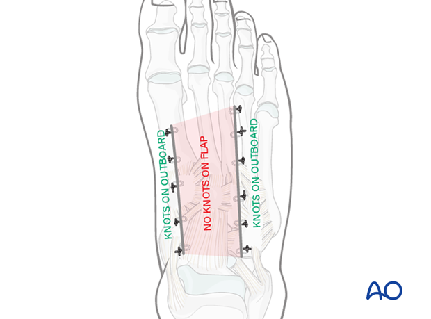
Percutaneous approach to the navicular
1. Indications
The percutaneous approach to the navicular can be used for screw fixation in non-displaced or minimally displaced simple/non-comminuted fractures (ie mid-body stress fractures).
2. Anatomy
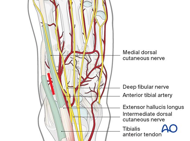
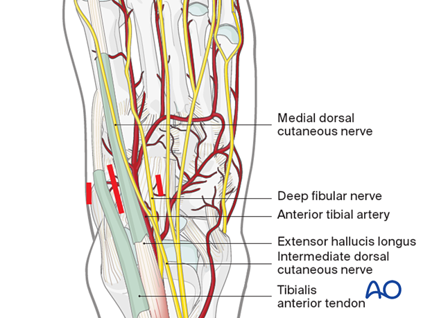
The dorsomedial approach to the navicular is made between the tibialis anterior tendon and the extensor hallucis longus (EHL) tendon. The approach should be made straight down from skin to periosteum without raising flaps or any unnecessary dissection.
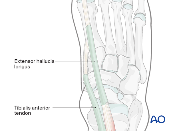
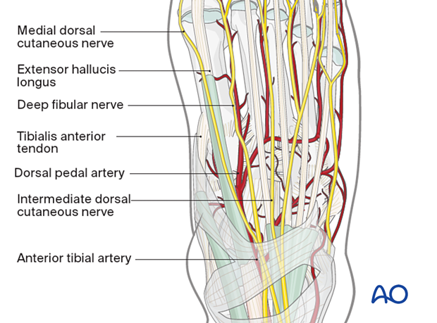
The neurovascular structures (dorsalis pedis artery) should be lateral to the EHL, but any small branches should be avoided or cauterized.
3. Skin incision
The full thickness incision uses the interval between the tibialis anterior and the EHL, directly over the fracture.

Two small stab incisions can be made dorsomedial and dorsolateral to allow for insertion of the pointed reduction clamps if needed.
4. Medial stab incision
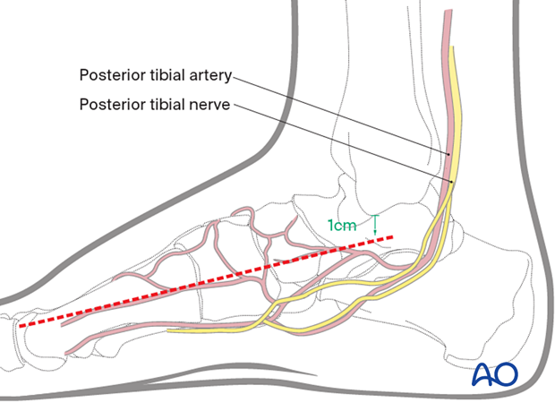
The stab incision medially is made in the line of the medial utility incision and could, if necessary, be enlarged.
Lag screws would then be applied through this incision, whatever its length.
5. Deep dissection
The fracture can be located using image intensification. The fracture site can then be directly entered. In the case of a non-healing stress fracture, the surfaces can be freshened by curetting or drilling. If a small cavity exists, this can be filled with a bone graft.
6. Wound closure
In general, wounds must be closed without tension on the skin edges.
The skin is closed without tension using an appropriate running everting suture (absorbable). In the case of multiple adjacent incisions (double dorsal Lisfranc approach) nylon can be used.
