Heterotopic ossification
1. Introduction
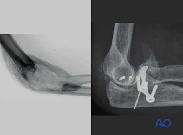
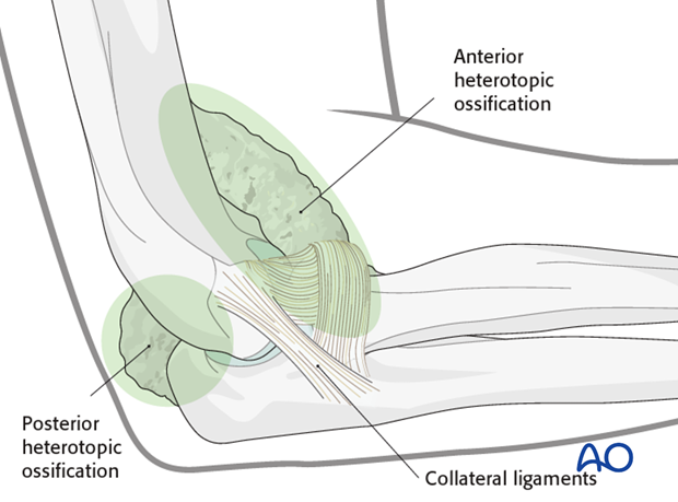
Formation of heterotopic bone (mostly in muscle) or peri-articular ossifications (around capsule and ligaments) around the elbow is common. It is a known sequela of elbow trauma (up to 37%), severe burns, or injury to the central nervous system. Severity ranges from minor clinically insignificant flecks of bone to complete bony ankylosis.
2. Etiology
Factors that contribute to heterotopic ossification include the complexity of fracture and/or dislocation, the extent of soft tissue injury, whether the fracture was open or not, surgical exposure and technique, and possibly timing of surgery.
3. Symptoms
Pain, edema, swelling and warmth may be more than expected as heterotopic ossification is forming in the first several weeks after insult, but generally becomes asymptomatic. The main symptom is loss of motion. Fortunately, most patients that develop only a small amount of heterotopic or peri-articular ossifications do not need surgery.
4. Diagnosis
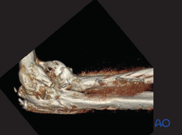
Radiographs and 3D CT. Pre-operative electrodiagnostic testing of ulnar nerve function can be helpful. There is no value in pre-operative bone scan, erythrocyte sedimentation rate (ESR), or alkaline phosphatase measurements.
Classification (Hastings) is based on location and/or functional impairment:
Class I - No functional limitation
Class II - Limitation in ≥ 1 plane
Class III - Ankylosis
Subdivision for II and III:
A (flex/ext)
B (pro/sup)
C (both)
5. Indication for surgical resection
Functional impairment and/or progressive ulnar nerve deficit. Timing of surgery has been debated, but heterotopic bone should be mature on radiographs. Early (4-6 months) is better as the patient is more motivated, the bone is softer, and there is less muscle atrophy. Contra-indications are infection, nonunion, and poor motor control.
6. Skin incision
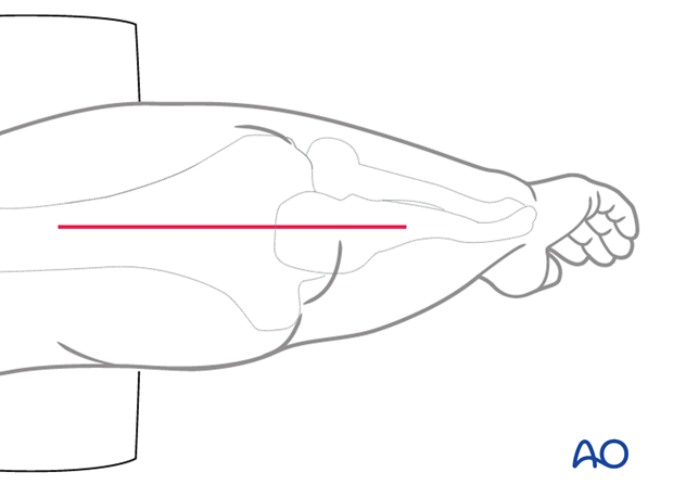
A posterior midline incision allows exposure of the medial, lateral, anterior and posterior elbow. The incisions might be dictated by previous incisions or skin quality (eg in patients with burns).
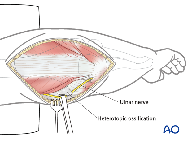
Identify the ulnar nerve first (pre-op CT and previous op-notes should be scrutinized for details of location of ulnar nerve) as the nerve may be encased in bone, particularly with heterotopic ossification associated with severe burns. It is often easiest to locate the nerve proximal and then trace it distally.
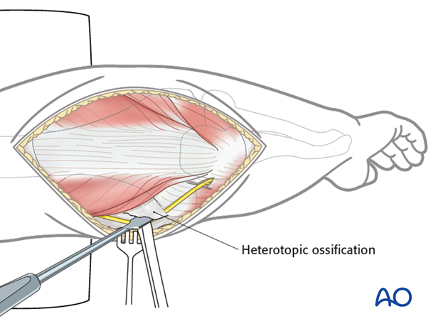
Excess bone around the nerve can be removed with a rongeur or small osteotome.
7. Lateral column procedure
If there is any chance the radial nerve or posterior interosseous nerve (PIN) are at risk, they need to be identified.
The extensor carpi radialis longus and brevis of the humerus along with brachialis are elevated.
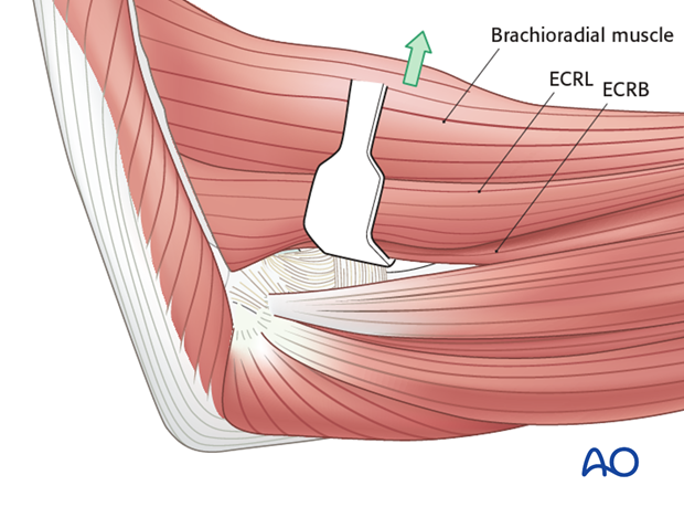
Open the interval between extensor carpi radialis brevis and extensor digitorum communis. The lateral collateral ligament should be spared.
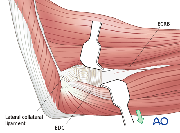
On the posterior side, the triceps and anconeus are elevated off of the distal humerus and ulna, respectively.
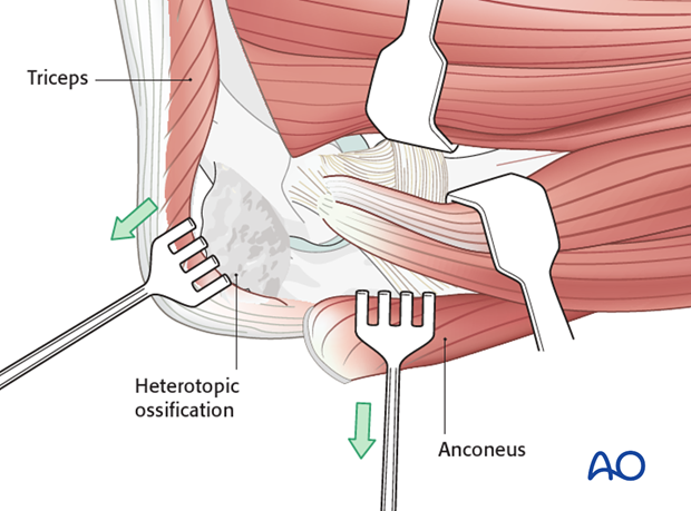
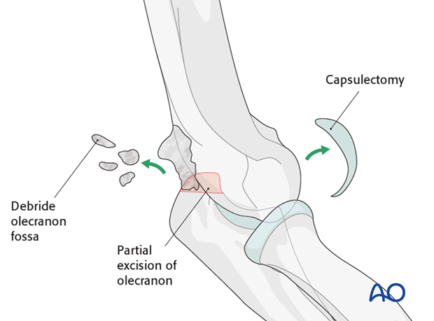
This approach will allow a capsulectomy and debridement of the coronoid fossa, olecranon tip, and olecranon fossa.
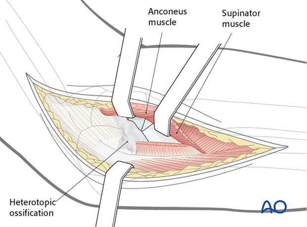
Heterotopic bone between the radius and ulna (or even a synostosis) can be resected using the Boyd approach elevating the extensor carpi ulnaris and supinator of the ulna.
8. Medial
The over-the-top approach elevates the brachialis muscle off the distal humerus along with the anterior half of the flexor-pronator mass that is divided horizontally.
The oblique band of the medial ligament (MCL) should be preserved whereas the posterior and transverse band of the MCL can be excised if needed.
Posteriorly the triceps is elevated off the distal humerus.
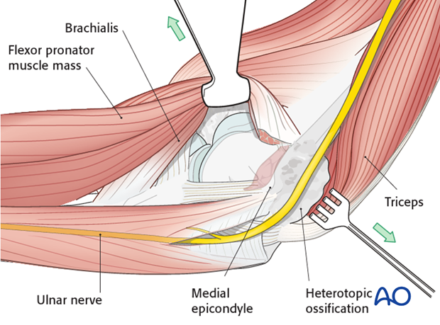
It is safer to leave all implants from osteosynthesis in place until manipulation is completed. Once heterotopic bone is removed, the elbow is gently mobilized to confirm restoration of motion.
If motion is still limited reassess adequacy of bone excision or consider additional soft tissue release (rarely the case). Forceful manipulation can cause refracture and must be avoided.
An ossified medial collateral ligament may fracture creating some instability on the medial side, but the elbow rarely subluxates or dislocates and no additional treatment of instability is needed.
9. Post-operative rehabilitation
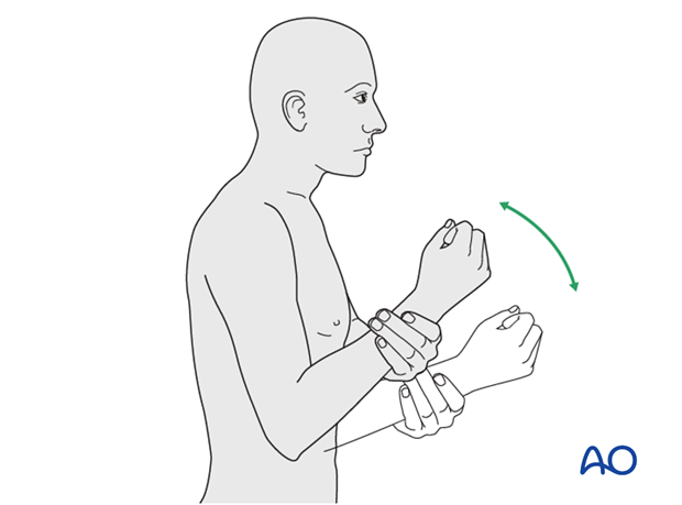
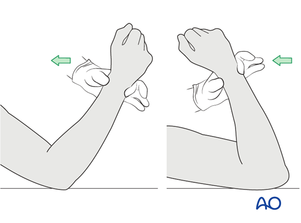
Active, self-assisted or gravity assisted ROM is started immediately. The use of a continuous passive motion machine (CPM) is optional. Immediately pre-or postoperatively a single dose of radiation (700 Gy) can be given to help prevent recurrence.
Results of heterotopic ossification resection are often quite good regardless of its cause and show worthwhile functional improvements despite risks of infection, recurrence, and nerve injury.
NOTE: There is NO role for radiation to prevent heterotopic ossification in fresh fractures where its use was shown to lead to nonunion.
