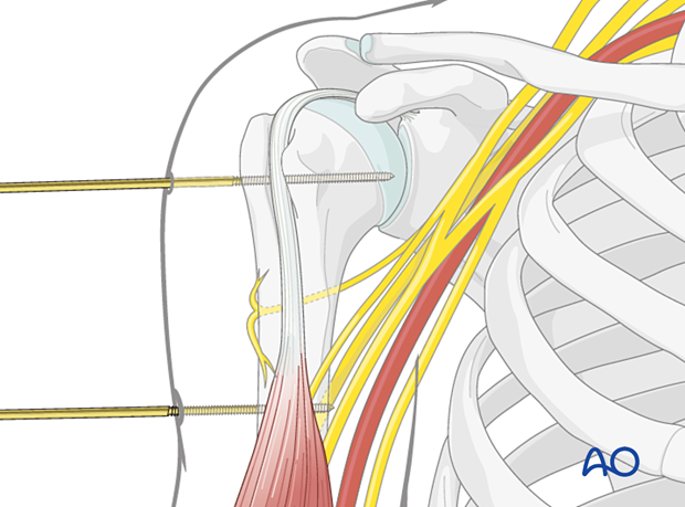
Safe zones in the humerus
1. Anatomy
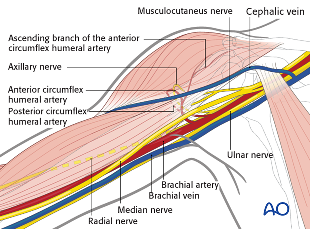
Neurovascular structures
The course of the following neurovascular structures should be kept in mind:
- Axillary nerve
- Cephalic vein
- Anterior circumflex humeral artery
- Ascending branch of the anterior circumflex humeral artery
- Posterior circumflex humeral artery
- Musculocutaneus nerve
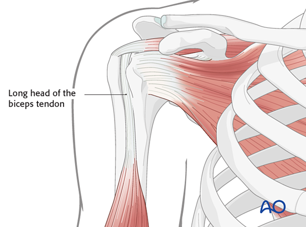
Tendon of the long head of the biceps
Another important structure is the tendinous origin of the long head of the biceps, which runs anteriorly and is usually palpable. Make sure not to fix this structure to the bone with pins or K-wires.
2. Safe zones
Axillary nerve
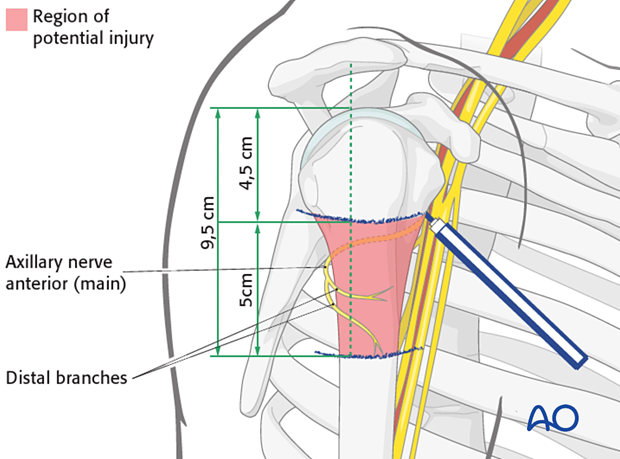
Anterior view of the proximal humerusThe anterior motor branch of the axillary nerve crosses the humerus horizontally about 6 cm distal to the cranial edge of the humeral head. This distance should not be considered an absolute guide for the actual position of the nerve or its branches: the position of the nerve varies considerably within the given limits, and therefore one should take this entire zone as a potential area for nerve damage.
The relationship between the main trunk and three terminal branches of the axillary nerve in relation to the top of the head of the humerus should also be taken into account, especially in a lateral approach, since they innervate one of the three deltoid muscle regions.
The region of potential injury to the axillary nerve and its branches is from 5 to 9.5 cm distal to the top of the humeral head.
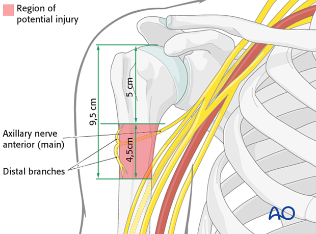
The region of potential injury to the axillary nerve and its branches is from 4.5 to 9.5 cm distal to the top of the humeral head.
3. Pin insertion
Safe zones for pins
Pins are inserted from lateral (alternatively from anterolateral or posterolateral) through the deltoid muscle.
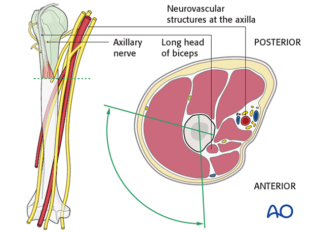
The tips of the pins in the humeral shaft should just perforate the far cortex. If inserted too deeply, they can injure the neurovascular bundle.
