Clinical evaluation
1. Overview
Fractures of the distal femur are rare in children.
Femoral fractures, especially in the nonwalking child, requires careful assessment of the circumstances of the injury and a whole-body physical examination to exclude deliberate injury.
2. Associated conditions
The fracture can involve the growth plate and/or the joint and this will influence the choice of treatment.
In rare cases these fractures may be associated with knee ligament injuries.
3. Symptoms and signs
- Thigh pain
- Knee pain
- Inability to walk
- Deformity of the injured leg
- Knee effusion
- Varus or valgus knee instability
4. Physical examination
Check for soft-tissue injuries and neurovascular compromise.
Check for injuries at other sites, especially in high-energy trauma, using standard assessment algorithms (ATLS).
Carefully examine for a vascular injury particularly after a high-energy mechanism and with significantly displaced fractures.
Twisting injuries
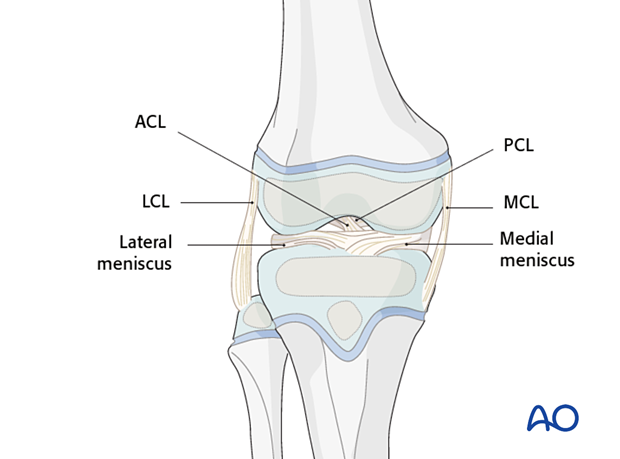
Twisting injuries may result in disruption of the medial or lateral collateral (MCL, LCL), anterior or posterior cruciate ligaments (ACL, PCL).
The MCL and ACL are most commonly injured.
Small avulsion fractures may represent markers of significant injuries, including knee dislocation and require MRI to determine the extent of the injury.

Sports trauma
Sports trauma can produce occult or undisplaced growth plate injuries. Careful clinical examination may identify tenderness and swelling at the growth plate.
Intraarticular fractures
Intraarticular fractures should be suspected if there is a significant hemarthrosis following a knee injury.
Aspiration of blood mixed with fat globules from the knee is indicative of an intraarticular fracture.
5. Vascular examination
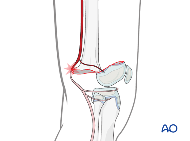
Vascular injury may occur with high-energy or displaced distal femoral fractures.
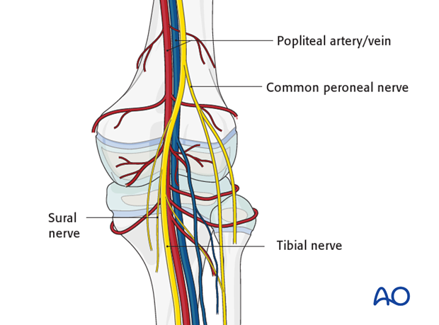
The popliteal artery lies directly behind the distal femoral metaphysis and is at particular risk in hyperextension injuries causing fractures with significant displacement.
Vascular consultation and arteriography is recommended in cases where vascular injury is suspected.
If delayed reperfusion has occurred the patient is also at risk of developing a compartment syndrome of the lower leg.

6. Neurological examination
The peroneal nerve can be injured by anterior and/or medial displacement of the distal fragment.
Immediate exploration is rarely necessary but may be considered if neurological loss persists and EMG studies suggest complete disruption.


