Ulnar approach to the pediatric distal ulna
1. Skin incision
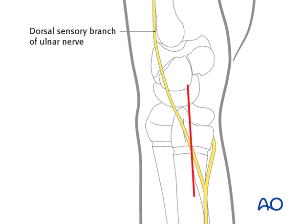
The ulnar shaft and the fracture gap between the ulnar styloid and the distal metaphysis are usually easily palpated.
A straight, longitudinal incision is made over the distal ulna, between the tendons of the extensor and flexor carpi ulnaris.

2. Surgical dissection
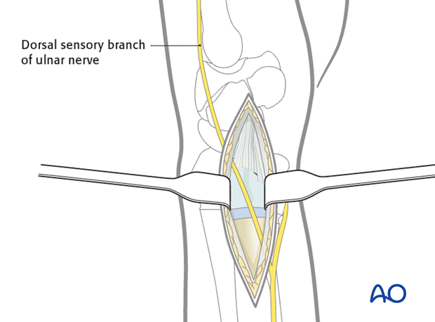
The dorsal sensory branch of the ulnar nerve must be sought and protected. Great care should be taken to avoid injury to this nerve.
The fracture site is then exposed, if necessary, releasing the ulnar attachment of the extensor retinaculum.

3. Wound closure
The extensor retinaculum is repaired, as necessary, and the wound is closed in layers, again taking care not to damage the dorsal sensory branch of the ulnar nerve.


