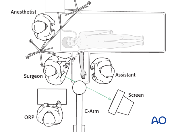
Supine positioning
1. Patient positioning
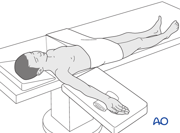
The patient is positioned supine and the forearm is placed on an arm table.
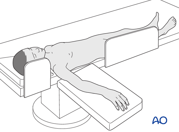
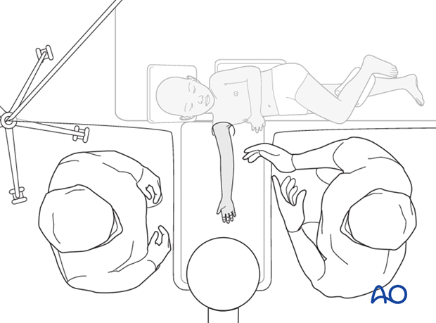
For small children, it may be necessary for the shoulder to rest on the arm table to allow unimpeded imaging. This may require gentle lateral flexion of the neck and trunk with appropriate padding and attention to pressure areas.
By abducting the shoulder it is possible for the surgeon and the assistant to sit on either side of the hand table.
A pneumatic tourniquet is used.
Prophylactic antibiotics are optional, according to local microbiological protocols.
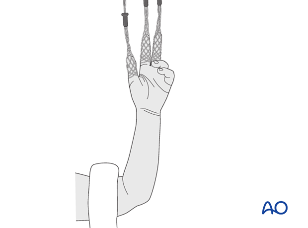
Finger traps can be used to apply traction and assist the closed reduction of some fractures.
A rolled up towel, or bag of saline, can be placed under the supinated distal forearm to improve exposure and allow dorsiflexion of the wrist.
2. OR set-up
The optimal position of the surgeon is defined by the anatomical requirements of the fixation. The position of the surgeon and assistant depends on forearm pronation/supination and which of the bones is involved (as shown below).
The position of the limb should allow complete imaging in the frontal and sagittal planes of the distal radius and/or ulna.
The position of the screen should allow a direct line of sight for the surgeon.
The position of operating room personnel should allow ergonomic assistance.
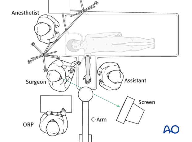
Depending on the anatomical requirement of the fixation, there are four possibilities for arranging the OR (right-hand-dominant surgeon illustrated):
1. Supinated forearm for treatment of isolated radial, or radial and ulnar, fractures

2. Supinated forearm for treatment of ulnar fractures
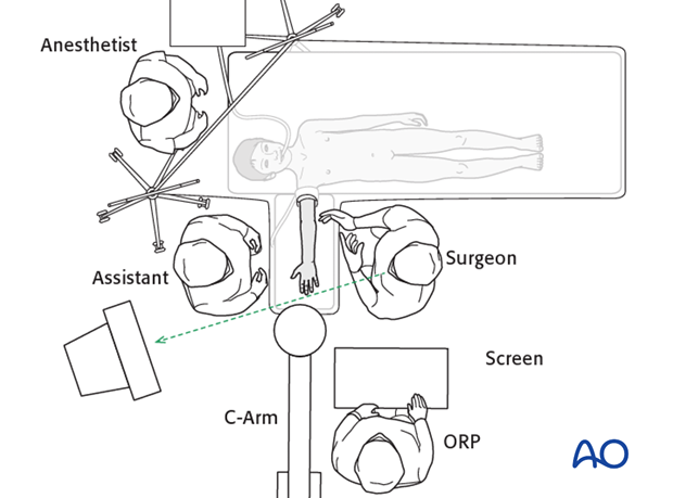
3. Pronated forearm for treatment of isolated radial, or radial and ulnar, fractures
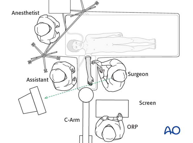
4. Pronated forearm for treatment of ulnar fractures
