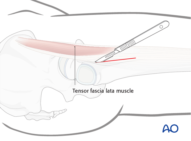
Anterior approach to the pediatric proximal femur
1. Preliminary remarks
The anterior approach provides the most direct access to the anterior aspect of the hip. Many surgeons prefer this approach for reduction of femoral head and neck fractures.
Note: Fixation of femoral neck fractures reduced through this approach will require separate percutaneous screw insertion, or a separate lateral incision.
2. Vascular anatomy
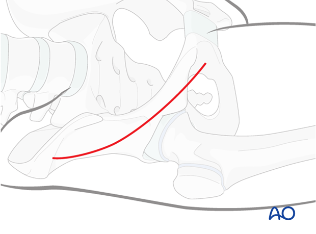
The deep branch of the medial femoral circumflex artery provides the main relevant blood supply to the femoral head.
The medial femoral circumflex artery originates from the deep femoral artery (profunda femoris), courses between the iliopsoas and pectineus muscles, and runs posteriorly between the femur and the pelvis.
During its course, a small branch supplies the inferior retinaculum (ligament of Weitbrecht).
The main branch of the medial femoral circumflex artery is related to the inferior border of the obturator externus muscle and passes posterior to the femur, towards the intertrochanteric crest.
It then crosses posterior to the obturator externus and anterior to the triceps coxae (obturator internus and the superior and inferior gemelli).
Before crossing the triceps coxae, a small branch passes to the greater trochanter.
The vessel enters the joint capsule between the gemellus superior and the piriformis muscles.
Note: The approach must always be cranial to the piriformis muscle. This anatomical detail is crucial when starting to prepare the capsule.
After perforating the capsule, the vessel passes along the superior retinaculum and splits into 3-4 branches.
Provided the obturator externus muscle remains intact, it will protect the medial femoral circumflex artery.
3. Skin incision
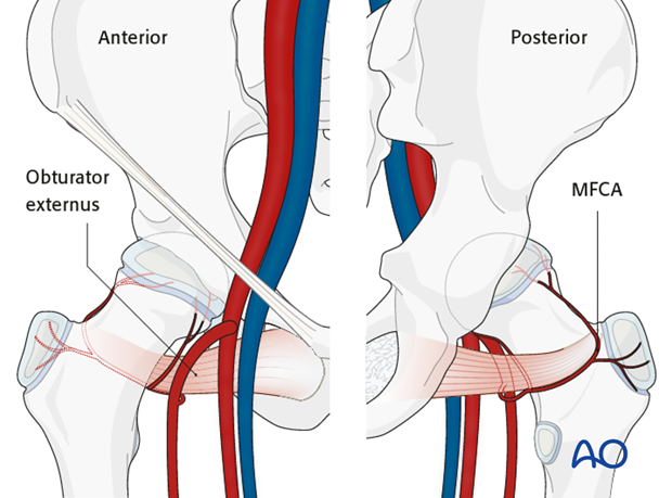
A bikini incision is used. The level of the incision is below the anterior superior iliac spine and centered on the anterior inferior iliac spine.

4. Deep dissection
After incision of the skin and fat, the deep fascia is encountered.
The interval between tensor fascia lata and sartorius is identified by palpation and the fascia incised.
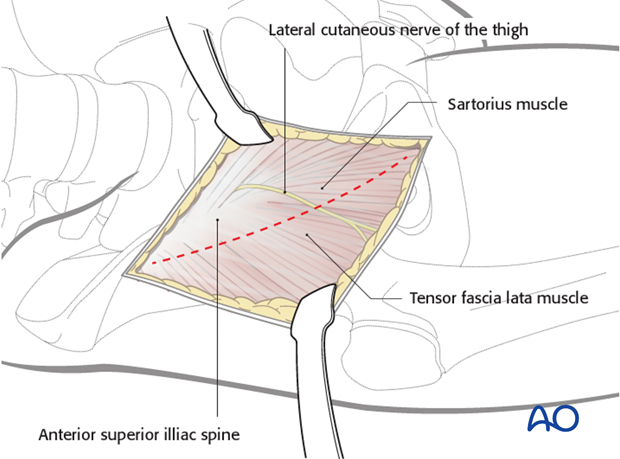
The fascia should be carefully incised and the lateral cutaneous nerve of the thigh identified and protected.
Note: Dissection within the medial edge of tensor fascia lata is preferred by some surgeons and helps to protect the lateral cutaneous nerve of the thigh.
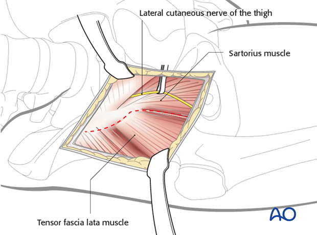
5. Development of interval between tensor fascia lata and sartorius
Having identified the nerve, the interval between tensor fascia lata and sartorius is opened.
In children, the muscles can frequently be adequately retracted without division of the apophysis of the iliac crest and retraction of the abductors. If access is restricted, the tensor fascia lata should be partially released from the iliac crest.
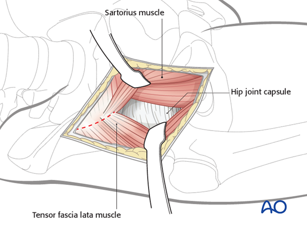
6. Deep surgical dissection
The straight head of rectus femoris is released from the anterior inferior iliac spine. This can be found by palpating down the ilium from the superior spine to the inferior spine.
The reflected head of rectus femoris is closely adherent to the capsule. This can be separated from the capsule, released and retracted.
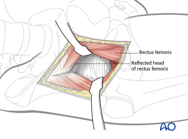
A Z-shaped capsulotomy, with stay sutures medially and laterally, allows exposure of the femoral head and neck. The labrum should be protected during the capsulotomy.
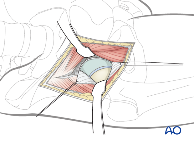
Screw, or plate, fixation of the fracture will require a separate lateral approach to the shaft, via either a stab, or a formal incision.
