Anterolateral approach to the pediatric proximal femur
1. Preliminary remarks
The anterolateral approach to the proximal femur, through the interval between the gluteus medius and minimus muscles and tensor fascia lata, provides access to the hip joint and the lateral proximal femur. This approach is adequate for fracture fixation and application of a plate onto the lateral aspect of the femur.
2. Vascular anatomy
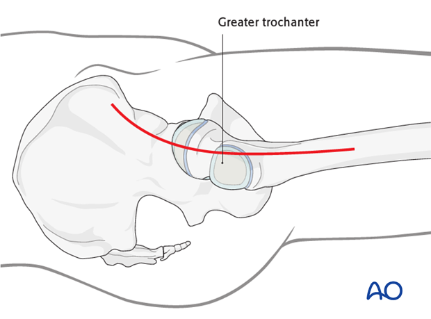
The deep branch of the medial femoral circumflex artery provides the main relevant blood supply to the femoral head.
The medial femoral circumflex artery originates from the deep femoral artery (profunda femoris), courses between the iliopsoas and pectineus muscles, and runs posteriorly between the femur and the pelvis.
During its course, a small branch supplies the inferior retinaculum (ligament of Weitbrecht).
The main branch of the medial femoral circumflex artery is related to the inferior border of the obturator externus muscle and passes posterior to the femur, towards the intertrochanteric crest.
It then crosses posterior to the obturator externus and anterior to the triceps coxae (obturator internus and the superior and inferior gemelli).
Before crossing the triceps coxae, a small branch passes to the greater trochanter.
The vessel enters the joint capsule between the gemellus superior and the piriformis muscles.
Note: The approach must always be cranial to the piriformis muscle. This anatomical detail is crucial when starting to prepare the capsule.
After perforating the capsule, the vessel passes along the superior retinaculum and splits into 3-4 branches.
Provided the obturator externus muscle remains intact, it will protect the medial femoral circumflex artery.
3. Skin incision
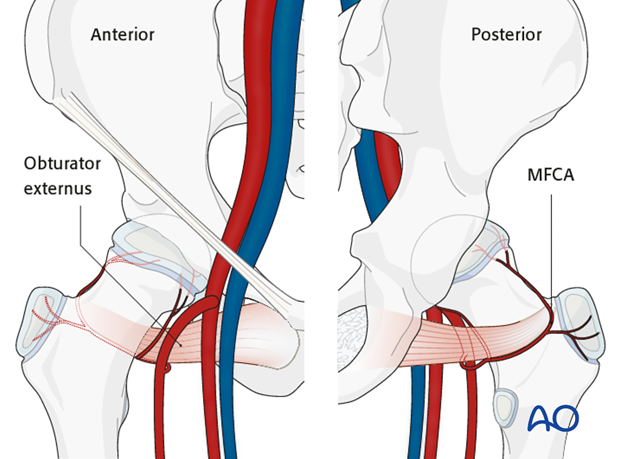
The incision starts just posterior to the anterior superior iliac spine, curves posteriorly and distally, crossing the anterior part of the greater trochanter, and continues down the line of the femoral shaft.

4. Incision of fascia lata
The skin and fat should be divided down to the deep fascia, which is cleared a little.
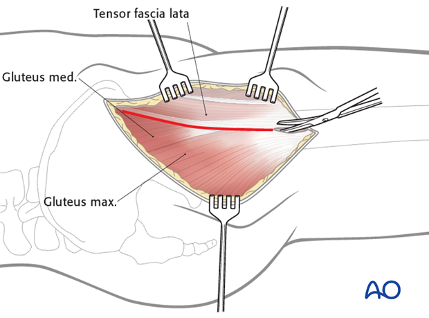
The tensor fascia lata sits within the fascia and the posterior edge of this muscle should be identified. The initial incision should be made in the region of the trochanter, or below, so that the anterior border of gluteus medius can be identified. The longitudinal fascia lata incision should be developed from distal to proximal, behind the tensor fascia lata muscle belly, remaining anterior to the gluteus medius.
5. Deep surgical dissection
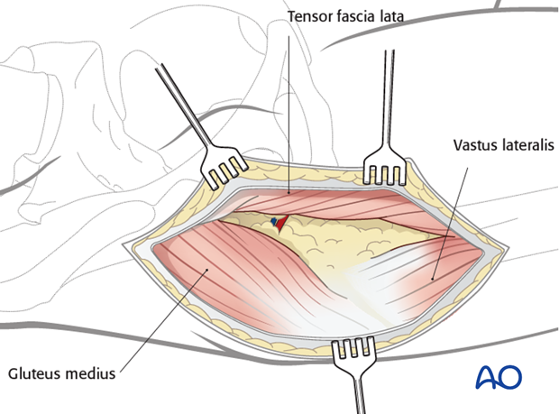
With the greater trochanter and the gluteus medius exposed, the tensor fascia lata is retracted anteriorly and the gluteus medius posteriorly. The interval between the gluteus medius and the tensor fascia lata is developed over the hip joint and extended proximally, by blunt dissection.
Be aware of vessels running across the proximal extent of this interval. They require ligation, or cautery.
The nerve to tensor fascia lata, found in the proximal part of the interval, should be preserved.
6. Exposure of the hip capsule
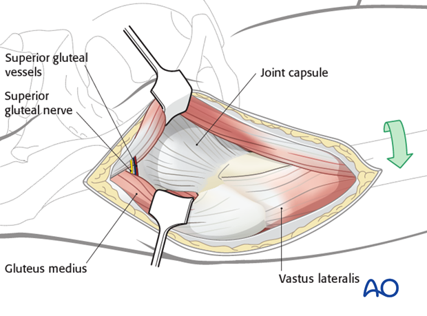
A retractor is placed anteriorly over the hip capsule, often onto the acetabulum. The fascia lata and tensor fascia lata are held open with a retractor. Additional anterior and posterior retractors will open the deeper interval.
External rotation of the leg improves access to the hip capsule.
7. Opening of the joint capsule
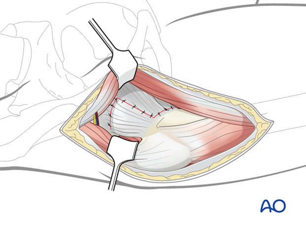
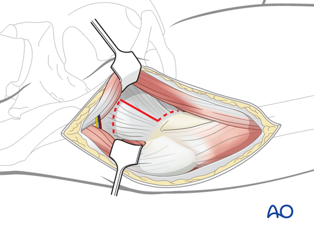
A longitudinal incision is made in the capsule. This can be extended inferiorly towards the lesser trochanter. A further extension can be made proximally, extending posteriorly, converting the capsulotomy to a Z.
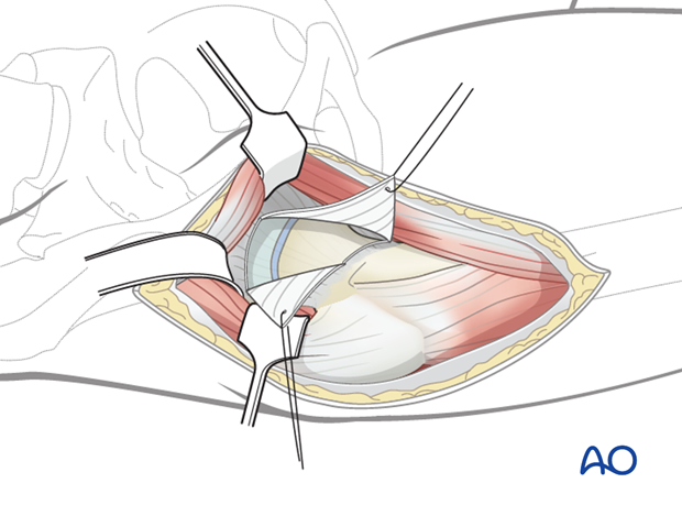
Two retraction sutures are inserted proximally and distally. The acetabular labrum is protected.
This capsulotomy exposes the anterior aspects of the femoral head and neck. Lateral traction and repositioning of the leg can improve visualization.
8. Release of vastus lateralis
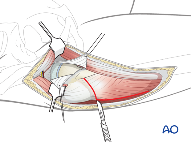
The incision can be extended distally over the proximal vastus lateralis to allow insertion of screws, or a proximal femoral plate, for femoral neck fracture fixation.
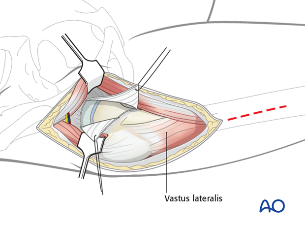
For plating, the vastus lateralis must be released from its origin. The muscle release needs to be limited anteriorly to protect the neurovascular supply. Posteriorly, the release can be as extensive as required.
Screw fixation can be performed without muscle release.
9. Wound closure
The capsule should be closed loosely to prevent the accumulation of a hemarthrosis under pressure. The fascia lata can be closed with a continuous suture.
The subcutaneous fat and the skin are closed according to the surgeon's preference.
