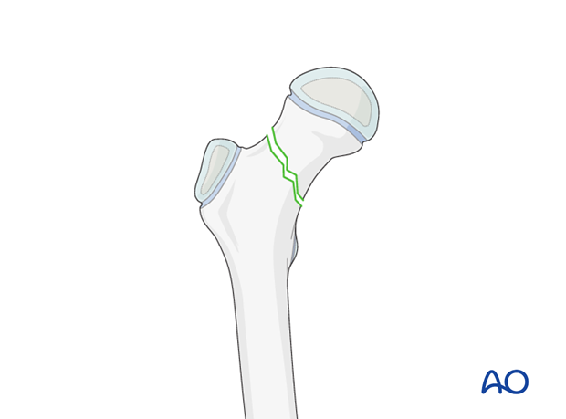
Open vs closed reduction
1. Introduction
Open reduction is used for the majority of pediatric femoral neck fractures.
Reasons for considering open reduction include:
- To reduce the risk of avascular necrosis
- To avoid/prevent the risk of delayed union/nonunion
- To avoid/prevent the risk of malunion
2. Avascular necrosis
Introduction
Avascular necrosis (AVN) is the most common serious complication of femoral neck fractures in children. Published literature estimates an overall risk of this complication of approximately 20%. Precise figures are, however, difficult to cite because of small numbers of patients in most series and considerable variation in treatment methods reported.
The risk of AVN may be influenced by:
- Mechanism of injury (high-energy vs low-energy fracture)
- Initial displacement of the fracture
- Fracture pattern
- Timing of reduction
- Possibility of damage to the blood supply during reduction maneuvers
- Quality of final reduction
- Stability of the fixation
- Decompression of the joint cavity at the time of surgery
Factors which are directly under the surgeon’s control include:
- Force applied during reduction
- Accuracy of reduction
- Decompression of hemarthrosis
- Stability of the fixation
Control of these factors may reduce the incidence of AVN, and, in many cases, will require open reduction and internal fixation.
In cases where in-situ fixation is performed, the joint cavity can safely be decompressed using a limited anterior approach.
Anatomy
Detailed knowledge of the anatomy of the proximal femoral blood supply is the key to successful management of femoral neck fractures.
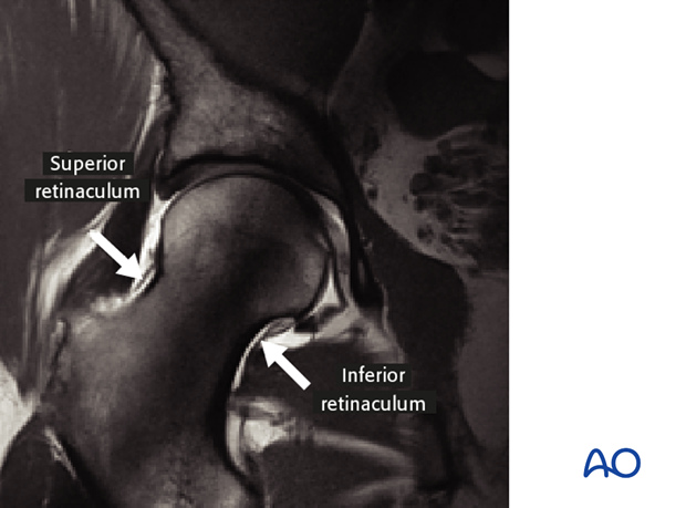
The two main vessels for femoral head blood supply run along the femoral neck within the inferior and superior retinacula. The strong structure of each retinaculum protects the vessels even when the fracture occurs.
Note: Incarceration of the superior retinaculum and the medial femoral circumflex artery in the fracture during closed reduction is an important cause of AVN.
The medial femoral circumflex artery
The deep branch of the medial femoral circumflex artery (MFCA) provides the main blood supply to the femoral head.
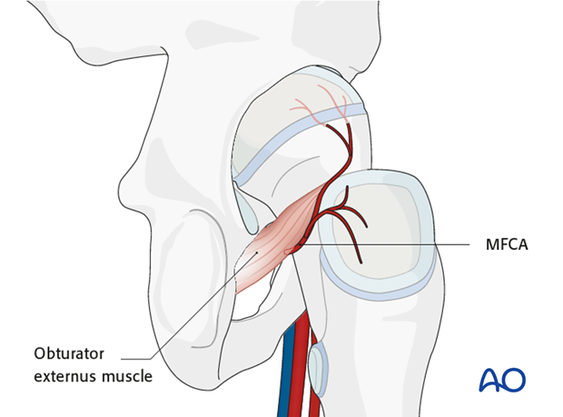
The MFCA arises from the profunda femoris artery anteriorly, and passes posteriorly, between iliopsoas and pectineus, to curve around the lower edge of the obturator externus muscle. It supplies the femoral head from posterior via the artery of the superior retinaculum.
Additionally, the MFCA also gives branches to the cruciate anastomosis that lies on the posterior surface of the quadratus femoris muscle.
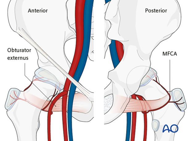
The branch within the inferior retinaculum (Weitbrecht's ligament) originates from MFCA.
The MFCA runs around the inferior border of the obturator externus muscle, as illustrated. This protects the MFCA from injury and it is essential to preserve this muscle and tendon.
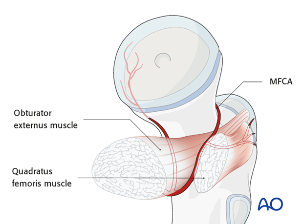
Note: The inferior and superior retinacula are only loosely attached to the periosteum of the femoral neck. This is demonstrated in the MRI with contrast. The vessels are therefore, not always damaged when the neck fracture displaces.

3. Closed reduction treatment with or without internal fixation
The following guidelines are the recommendations of the of AO Pediatric Expert Group, and are based on analysis of the contemporary literature.
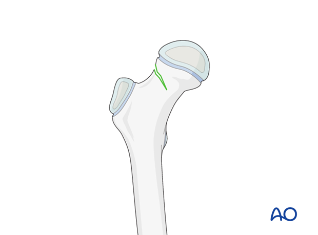
Unstable, complete fractures, with or without displacement
Complete, nondisplaced fractures are unstable and carry a high risk of secondary displacement, in addition to tension hemarthrosis.
It is, therefore, recommended to stabilize these fracture via an open approach to the hip.
Complete, displaced fractures should always be reduced and stabilized through an open approach, provided suitable expertise and facilities exist.
Closed management should be avoided.
