Lateral decubitus position
1. General considerations
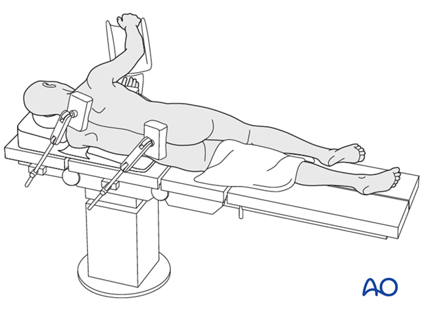
The patient is placed in a lateral decubitus position with the injured side up on either a radiolucent or on a fracture table which allows limb mobility.
Careful pre-cleaning of the soft tissues should be performed especially if gross contamination occurs.
Obese patients
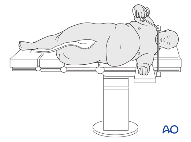
This position is recommended for obese patients.
In the lateral position, the fatty tissue falls away from the surgical field. This allows for proper visualization of the fracture and hemostasis.
Note: Meticulous soft tissue handling is particularly important in the medically infirmed elderly and obese patients, as a complication related to a periprosthetic femur fracture can lead to disastrous complications of the arthroplasty.
2. Preoperative preparation
Operating room personnel (ORP) need to know and confirm:
- Site and side of fracture
- Type of operation planned
- Ensure that operative site has been marked by the surgeon
- Condition of the soft tissues
- Implant to be used
- Patient positioning
- Details of the patient (including a signed consent form and appropriate antibiotic and thromboprophylaxis)
- Comorbidities, including allergies
3. Anesthesia
This procedure is performed with the patient under general or regional anesthesia.
4. Positioning
- The patient is supported in the lateral decubitus position on a radiolucent flat-top operating table.
- Pad all bony prominences and neurovascular structures carefully.
- A gel roll should be placed under the mid portion of the chest to protect the axilla and allow for ventilation
- Place the ipsilateral arm across the chest to be out of way.
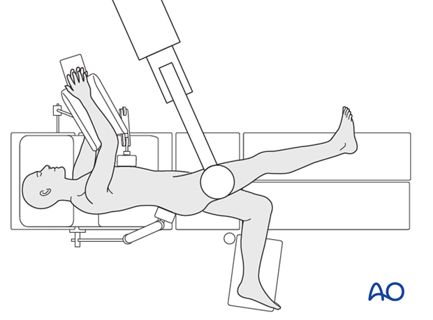
- Position the image intensifier on the opposite side of the injury and the operating surgeon.

- Ensure that you can get good-quality AP (illustrated) and lateral x-ray views of the fracture site, full prosthesis, and distal femur before draping.
5. Skin disinfecting and draping
- Disinfect the exposed area from above the iliac crest to the mid-tibia with the appropriate antiseptic.
- Ensure the adhesive portion of the drape is large enough to reach from the iliac crest to the knee joint
- Place the image intensifier on the nonsterile side of the exclusion drape.
- Traditional drapes may be used. Ensure a waterproof environment for the operative site.
- Drape the image intensifier.
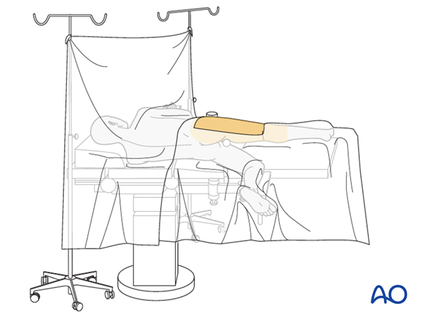
6. Operating room set-up
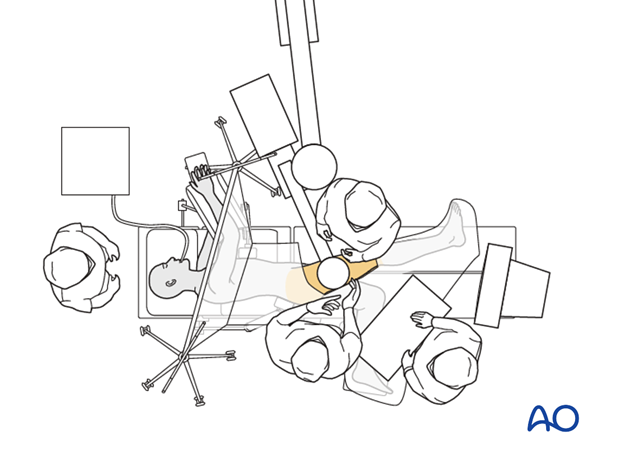
- Position the operating table (if feasible) within the operating room to allow maximum space on the operating side for the surgeon, staff, and trolleys.
- The surgeon and the ORP stand on the side of the injury. The Assistant may be on either side.
- Place the image intensifier on the opposite side of the patient, perpendicular to the patient.
- Place the image intensifier display screen in full view of the surgical team and the radiographer
Note: for periprosthetic fractures, whether performing ORIF or revision arthroplasty, it is preferred to position the patient on a radiolucent table.
