Supine position
1. Introduction
Variations of this position are useful for plating and nailing the distal femur, plate fixation of the proximal tibia, and fixation of the patella.
2. Preoperative preparation
- Verify site and side of fracture with the patient along with the type of operation planned
- Ensure that the operative site has been marked by the surgeon
- Condition of the soft tissues (fracture open or closed)
- Implant to be used (note: plate comes in right and left versions)
- Patient positioning
- Details of the patient (including a signed consent form and appropriate antibiotic and thromboprophylaxis)
- Comorbidities, including allergies
3. Anesthesia
This procedure is performed with the patient under general or regional anesthesia.
If a spinal anesthetic is used, the surgeon and anesthetist need to be confident that the procedure will not last more than 1.5 hours.
4. Prophylactic antibiotics
Antibiotics are administered according to local antibiotic policy and specific patient requirements.
5. Patient and x-ray positioning
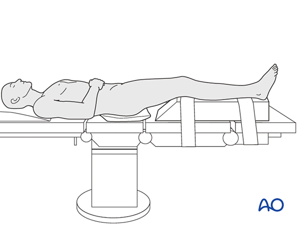
- Place the patient supine on a radiolucent operating table.
- Carefully pad all pressure points, especially in the elderly.
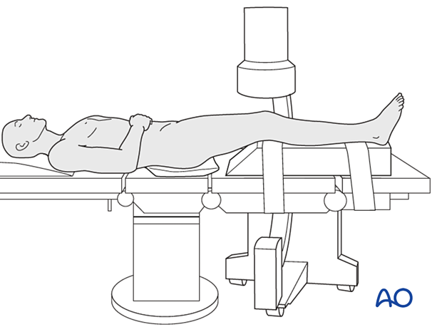
- Position the image intensifier on the opposite side of the injury and the surgeon.
- Before preparing and draping, ensure adequate imaging of the femur from the hip joint to the knee in the AP and lateral views.
- The C-arm should be able to pass freely under the table to obtain a cross-table lateral view in addition to the illustrated position for the AP view.
6. Skin disinfecting and draping
Maintain light manual traction (the assistant may need to stand on a stool) on the limb during preparation to avoid excessive deformity at the fracture site.
Disinfect the whole leg from the hip including the foot with the appropriate antiseptic.
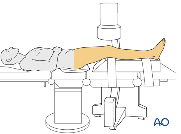
- Drape the limb with a single-use U-drape. A stockinette covers the lower leg and is fixed with tape.
- Consider prepping and draping the uninjured leg to facilitate comparison of frontal plane alignment and rotation
- Drape the image intensifier.
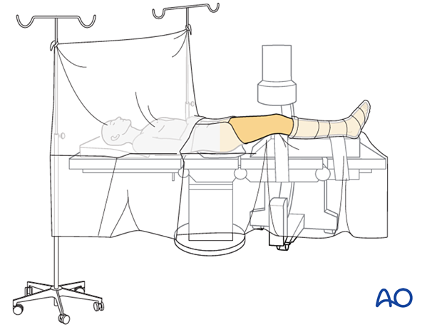
7. Operating room set-up
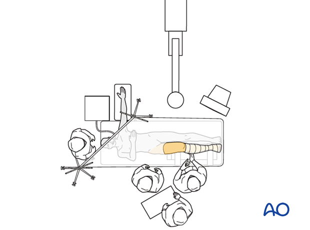
The surgeon and ORP stand on the side of the affected limb. The assistant stands next to the surgeon.
Place the image intensifier on the opposite side of the injury and the display screen in full view of the surgical team and the radiographer.
