Lateral approach to the zygomatic arch
1. Indications
The lateral approach to the zygomatic arch and ventral orbit are indicated for open reduction and internal fixation of zygomatic arch and orbital fractures.
2. Skin incision
The zygomatic arch and the orbit are identified by palpation. A skin incision is performed with a scalpel blade along the dorsal (a) or ventral (b) margins of the bone, and extended as needed to expose the fracture. The use of electrocautery should be avoided.
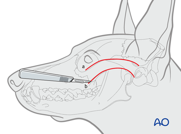
A standard approach to the fracture should always be used even when there are wounds in the area. Ideally, avoid going directly through the traumatic wound to prevent unnecessary trauma to the injured area. If the wounds are at the location of the standard approach, then going through the wounds may be necessary.
The exposure should be large enough for proper inspection and reduction of the fracture. The wound must be debrided gently, and vital tissues preserved.
3. Exposure
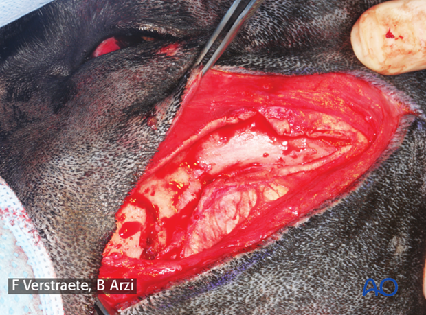
After the skin incision, the platysma muscles are incised. The periosteum, with origin on the masseter muscle, is incised along the ventral margin. The periosteum, with insertion on the temporal muscle along the dorsal margin, is also incised.
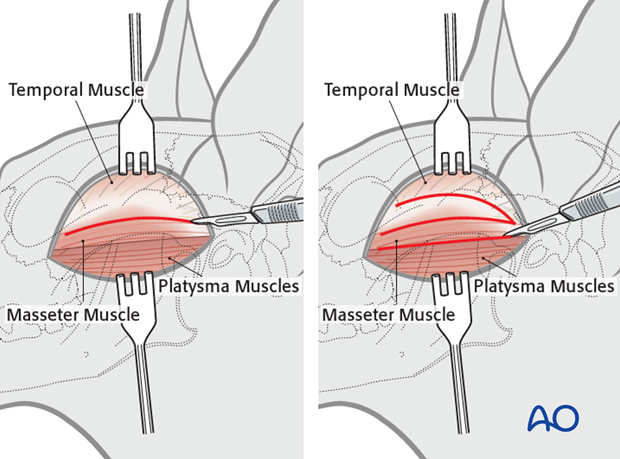
The tissues are elevated and retracted with the help of periosteal elevators.
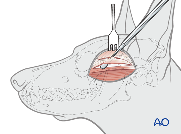
The medial aspect of the zygomatic arch is separated from its muscular attachments using periosteal elevators.
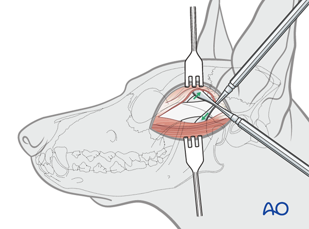
The image shows the elevated platysma and the incised periosteum, exposing the zygomatic arch fractures.
4. Closure
Closure is done in three layers. The first layer is the periosteum and muscles, then the platysma and subcutaneous tissue, followed by the skin. Closure of the first two layers is done with absorbable sutures such as 4.0 polyglactin 910 or poliglecaprone 25. The skin is closed with non-absorbable sutures, such as nylon, in a simple-interrupted fashion.
The use of staples should be avoided in the maxillofacial region because they can be irritating and can cause scaring in this area.


