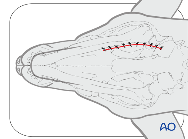
Ventral approach to the caudal mandible
1. Skin incision
With the patient in dorsal recumbency, the head is supported with a soft pad and the neck extended. The mandible is palpated, and the incision is planned just medial to the mandibular body. A skin incision is performed with a scalpel blade along the ventral medial margins of the bone and extended to include the angular process of the mandible. If both mandibles need to be exposed, two separate incisions may be needed.
The use of electrocautery should be avoided.
Note: The ventral border and the angular process of the mandible are palpated for surgical orientation. In severely displaced fractures, this may not be readily palpable.
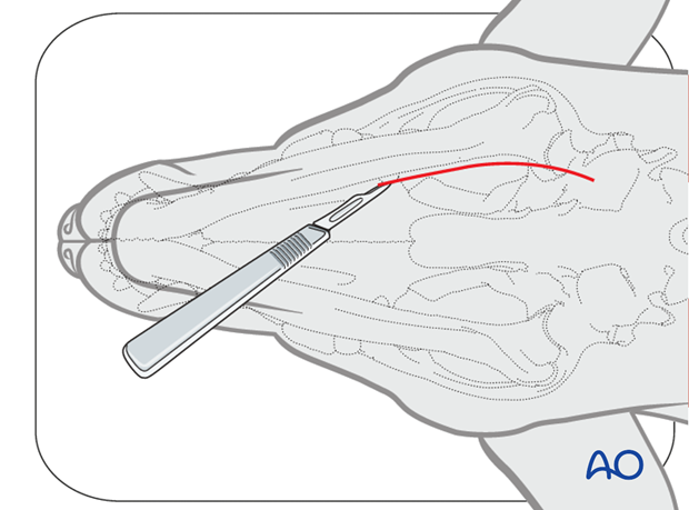
The approach should not be directly over the traumatic wound to avoid unnecessary trauma to the injured area. The exposure should be of sufficient size, so that the surgeon can inspect the wound and reduce the fracture without additional trauma to soft tissue.
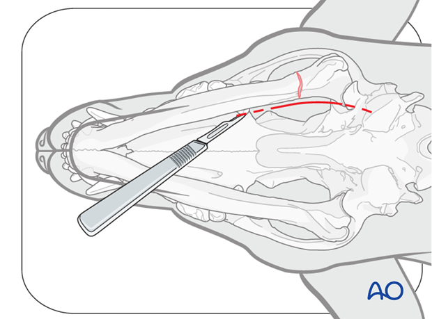
2. Exposure
The subcutaneous and platysma muscle are incised and retracted.
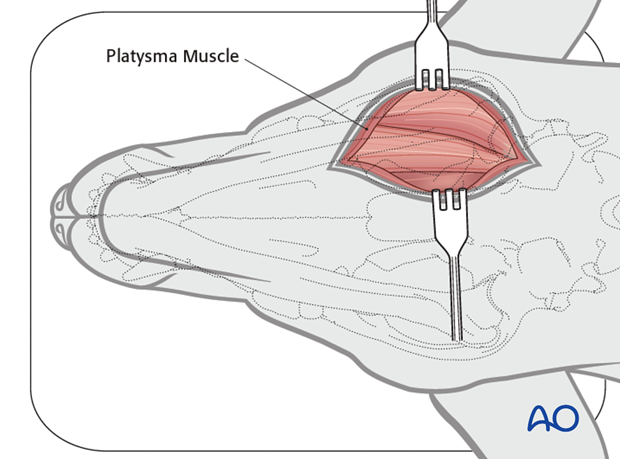
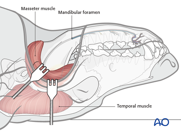
The rostral belly of the digastricus muscle and the masseter muscle are identified. The intermuscular septum is divided by blunt dissection until the periosteum and insertion of the masseteric fascia are reached.
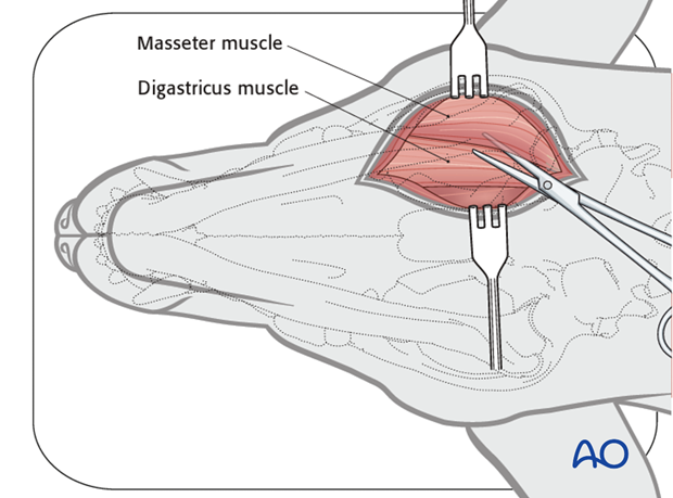
The periosteum is incised, and the two muscles are subperiosteally elevated and retracted.
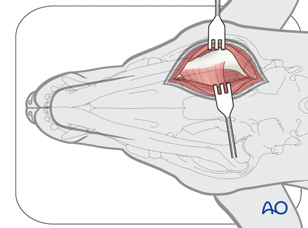
The insertion of the medial pterygoid muscle can be elevated to reach the most caudal aspect of the mandible.
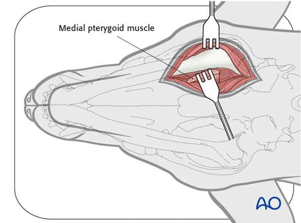
Care should be taken to avoid damaging the sublingual artery, branches of the facial artery and branches of the facial veins. Identification and isolation of the lingual nerve is important.
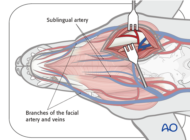
The masseter muscle is elevated from the masseteric fossa dorsally. Care should be taken to avoid the mandibular foramen and its associated neurovascular bundle during medial exposure.
3. Closure
Closure is done in three layers. The first layer is the periosteum and elevated muscles, then the platysma and subcutaneous tissue, followed by the skin.
Closure of the first two layers is done with absorbable sutures such as 4.0 polyglactin 910 or poliglecaprone 25. The skin is closed with monofilament nonabsorbable sutures in a simple-interrupted fashion.
